The analysis of this case is also useful for understanding what happens during the first weeks after a rhinoseptoplasty. All postoperative photographs were taken one month after surgery: despite the complexity of the procedure, which required multiple osteotomies and several grafts, the definitive shape is already clearly evident, bruising is entirely absent, and the edema (postoperative swelling) has resolved by approximately 70–80%. Over the next months, the shape will continue to refine, albeit very slowly, as the remaining edema subsides, with complete resolution around 9–12 months after surgery.
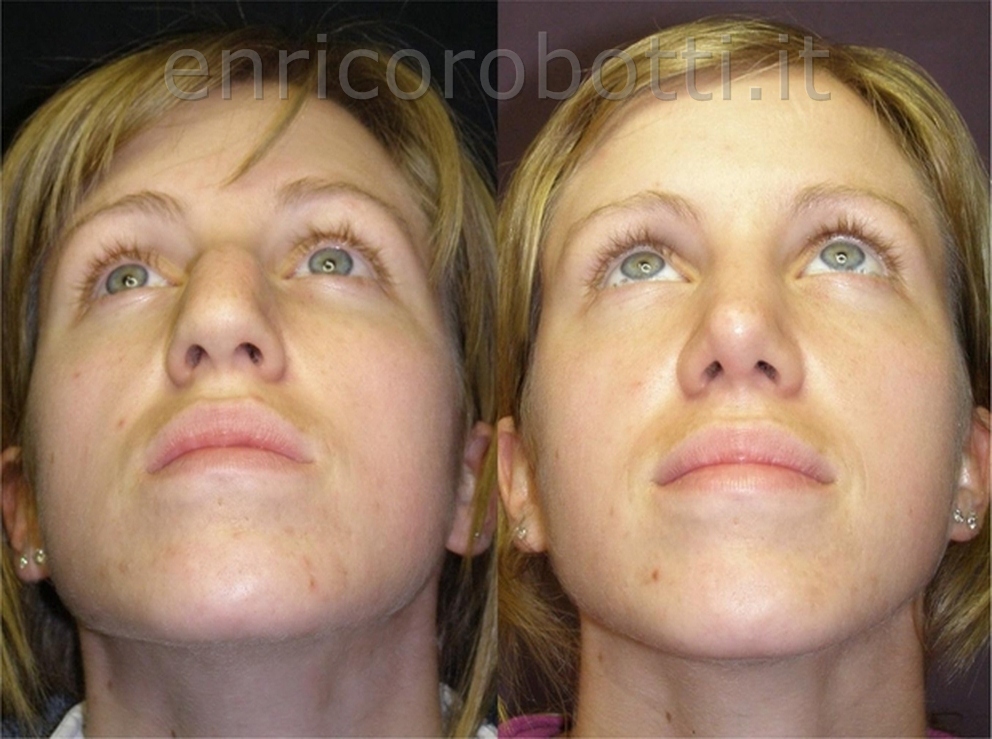
In this young woman, the nose is wide and deviated. The excessive width is primarily due to the nasal bones, which are too strong and overly separated. The deviation is both bony and cartilaginous, meaning it is caused by asymmetrical nasal bones and also (and mainly) by the nasal septum, which deviates to the right beginning at the point where the nasal bones end. Furthermore, the tip is rather bulbous and, above all, poorly supported. This becomes particularly evident when the patient smiles. If one were to treat only the dorsal hump (as might seem logical after a superficial assessment), the tip would droop even further downward, because it would lose the compensatory element that partially supports it—namely, the hump itself.
It is therefore clear that the hump must indeed be reduced, but, above all, the tip must be structurally supported and given a more sculpted shape through several grafts (to the columella and to the alar cartilages) harvested from the septum. This must be done before the hump is fully reduced; otherwise, the outcome would be a saddle deformity. Once again, this comes back to the fundamental concept of proportions in rhinoplasty, achieved through a carefully balanced combination of reduction (of excessive components) and augmentation (of deficient ones), reconstructing a stable long-term framework with appropriate use of various grafts.
As always, function and aesthetics are closely interrelated: in this case, it is primarily the deviated septum that causes the breathing impairment, as confirmed by the CT scan, which is always advisable before surgery. Complete mobilization and correction of the septum, along with the correction of the nasal bones, will restore the aesthetic lines of the dorsum and correct the respiratory deficit—more than compensating for the inevitable narrowing of the airway that follows osteotomy and medialization of nasal bones that are excessively wide. Put differently, since it will be necessary to reposition the nasal bones closer together to achieve proper aesthetic dorsal lines, if this were done without completely correcting the septal deviation, the respiratory function—which is already compromised preoperatively—would become significantly worse after surgery.