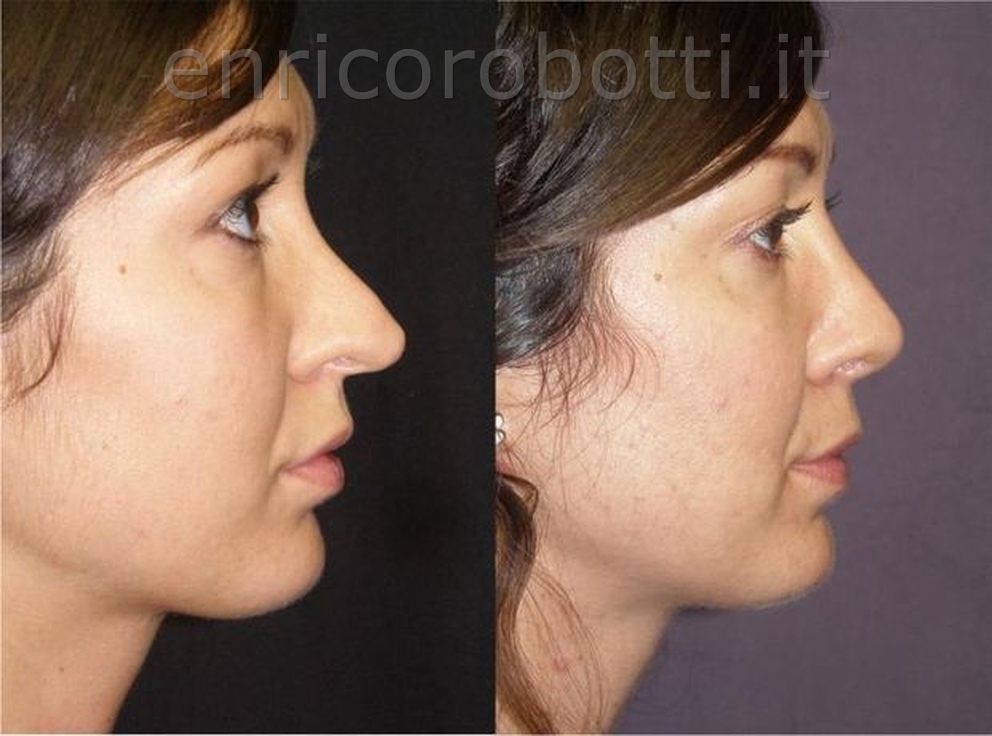
In this patient, who had previously undergone a closed technique, an inverted-V deformity of the middle third of the dorsum is evident on frontal view, due to collapse of the upper lateral (triangular) cartilages. The tip appears irregular, with asymmetry of the two domes, and is also long and overprojected. On profile view, both the saddle deformity of the dorsum, due to excessive dorsal hump reduction, and the disharmony with the tip are evident. Here again, the goal will be to restore correct proportions by re-shaping the structures that were not adequately treated (the tip cartilages) and by restoring, with grafts, the portion of the dorsum that was excessively removed. The alar cartilages will be shortened and symmetrized. The two domes, which had been sutured to each other, creating an inappropriate “single tip” appearance, will be separated and positioned at the correct distance from one another. After shortening, the tip will be slightly rotated upward and stabilized with a septal extension graft harvested from the proximal portion of the septum. As for the dorsum, the inverted-V deformity will be corrected with spreader grafts, also harvested from the septum. The dorsum will then be augmented with diced cartilage wrapped in temporalis fascia (“diced cartilage fascia”). Respiratory function, which was completely inadequate, is corrected in this patient both by septoplasty of the residual obstructing portion of the septum and by opening the internal nasal valve with spreader grafts. In this case as well, as in every primary or revision rhinoplasty, Dr. Robotti avoided the use of any alloplastic material, employing only grafts harvested from the patient herself. Fortunately, the septum had not been manipulated previously and therefore provided a sufficient amount of cartilage for these grafts.